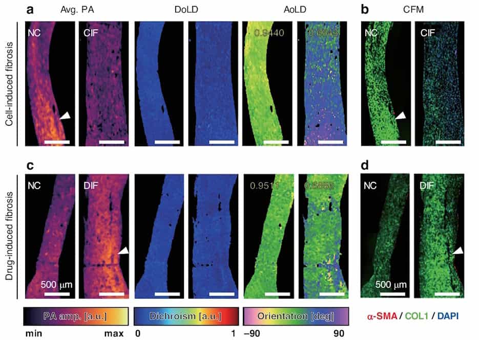
Imaging tissue fibrosis (a) MIR-DS-PAM images of cell-induced fibrosis (CIF) and normal control (NC) tissue; (c) MIR-DS-PAM images of drug-induced fibrosis (DIF) and NC tissue; (b) and (d) show the corresponding confocal fluorescence microscopy (CFM) images. Scale bars: 500 μm. (Courtesy: CC-BY 4.0/ Light Sci. Appl. 10.1038/s41377-025-02117-0)" title="Click to open image in popup" href="https://physicsworld.com/wp-content/uploads/2026/01/19-01-26-photoacoustic-microscopy-fig4.jpg"> Imaging tissue fibrosis (a) Mid-infrared dichroism-sensitive photoacoustic microscopy (MIR-DS-PAM) images of cell-induced fibrosis (CIF) and normal control (NC) tissue; (c) MIR-DS-PAM images of drug-induced fibrosis (DIF) and NC tissue; (b) and (d) show the corresponding confocal fluorescence microscopy (CFM) images. Scale bars: 500 µm. (Courtesy: CC-BY 4.0/ Light Sci. Appl. 10.1038/s41377-025-02117-0) Many of the tissues in the human body rely upon highly organized microstructures to function effectively.…