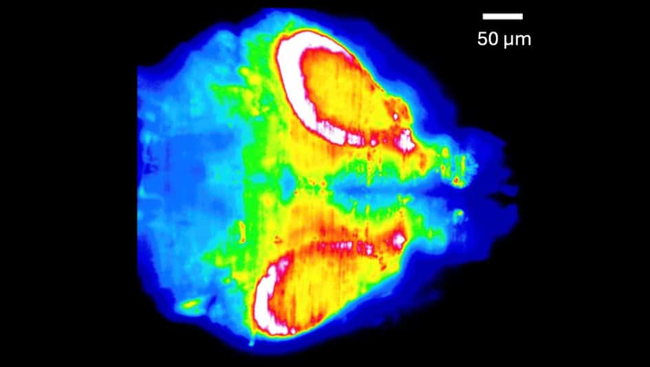
Mid-seizure activity Scientists capture a snapshot of a larval zebrafish brain during a seizure. White and red mark regions with the greatest neuronal activity, while blue tones indicate lower activity. (Courtesy: Adapted with permission from B Liu et al. Biomedical Optics Express 10.1364/BOE.596096 2026 © Optica Publishing Group)"> Mid-seizure activity Scientists capture a snapshot of a larval zebrafish brain during a seizure. White and red mark regions with the greatest neuronal activity, while blue tones indicate lower activity. (Courtesy: B Liu et al. Biomedical Optics Express 10.1364/BOE.596096 2026 © Optica Publishing Group)
Neuroscientists are debating a seemingly simple question: which way does a seizure propagate in the brain? Answering such a question could advance our understanding of epilepsy, but requires the development of cutting-edge microscopy techniques that are far from simple.…