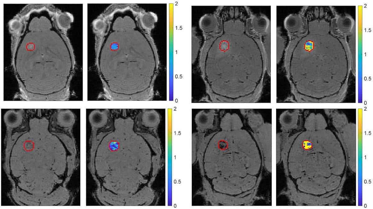
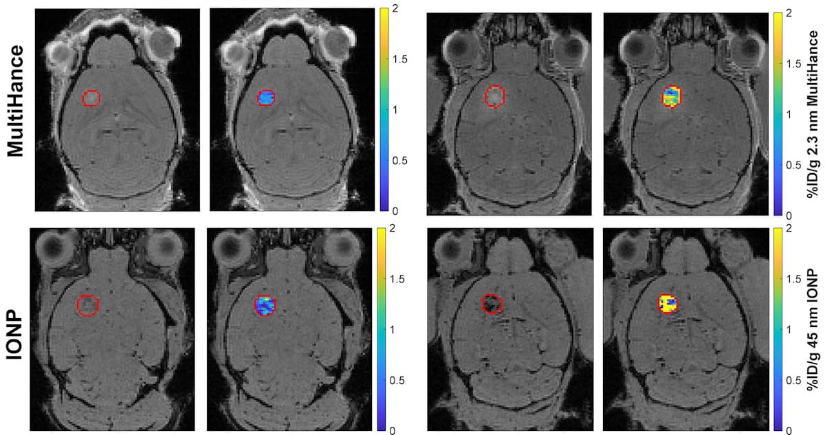
Brain tumour images Representative MR images of baseline (left panels) and FUS-treated (right panels) mice with tumour-bearing brains. The top row shows images for administration of a 2.3 nm contrast agent (MultiHance), the bottom row shows administered iron oxide nanoparticles, 45 nm in size. Each pair of panels shows the anatomic image with the tumour outlined in red on the left, and the corresponding image superimposed with a concentration map showing the amount of nanoparticle retained within the tumour (as a percentage of the injected dose per gram) on the right. (Courtesy: CC BY 4.0/ Radiology 10.1148/radiol.251005)" title="Click to open image in popup" href="https://physicsworld.com/wp-content/uploads/2026/09/1-09-26-fus-bbb-fig5.jpg"> Brain tumour images Representative MR images of baseline (left panels) and FUS-treated (right panels) mouse brains. The top row shows administration of a 2.3 nm contrast agent (MultiHance), the bottom row shows administered iron oxide nanoparticles, 45 nm in size.…