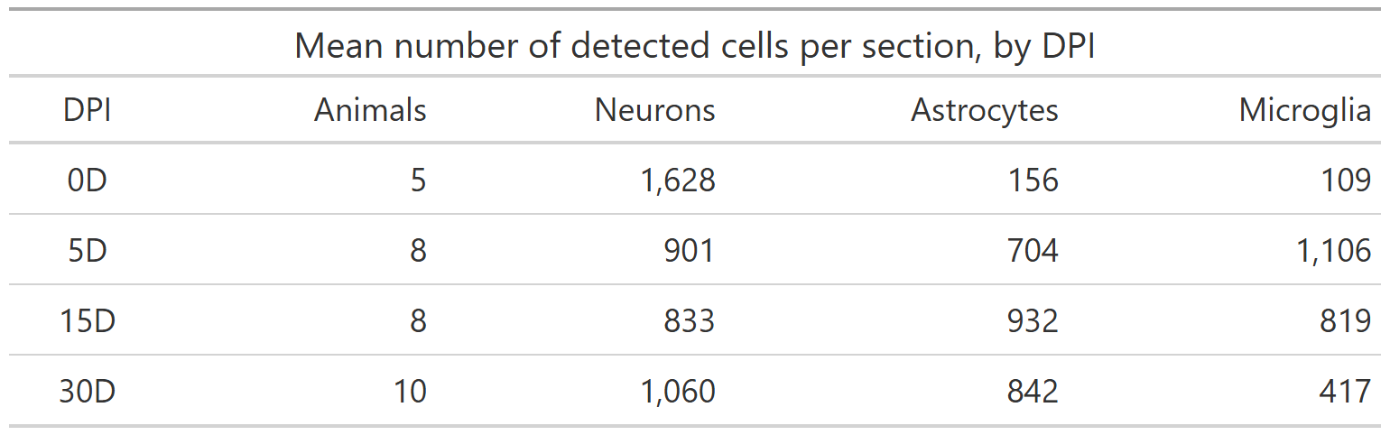
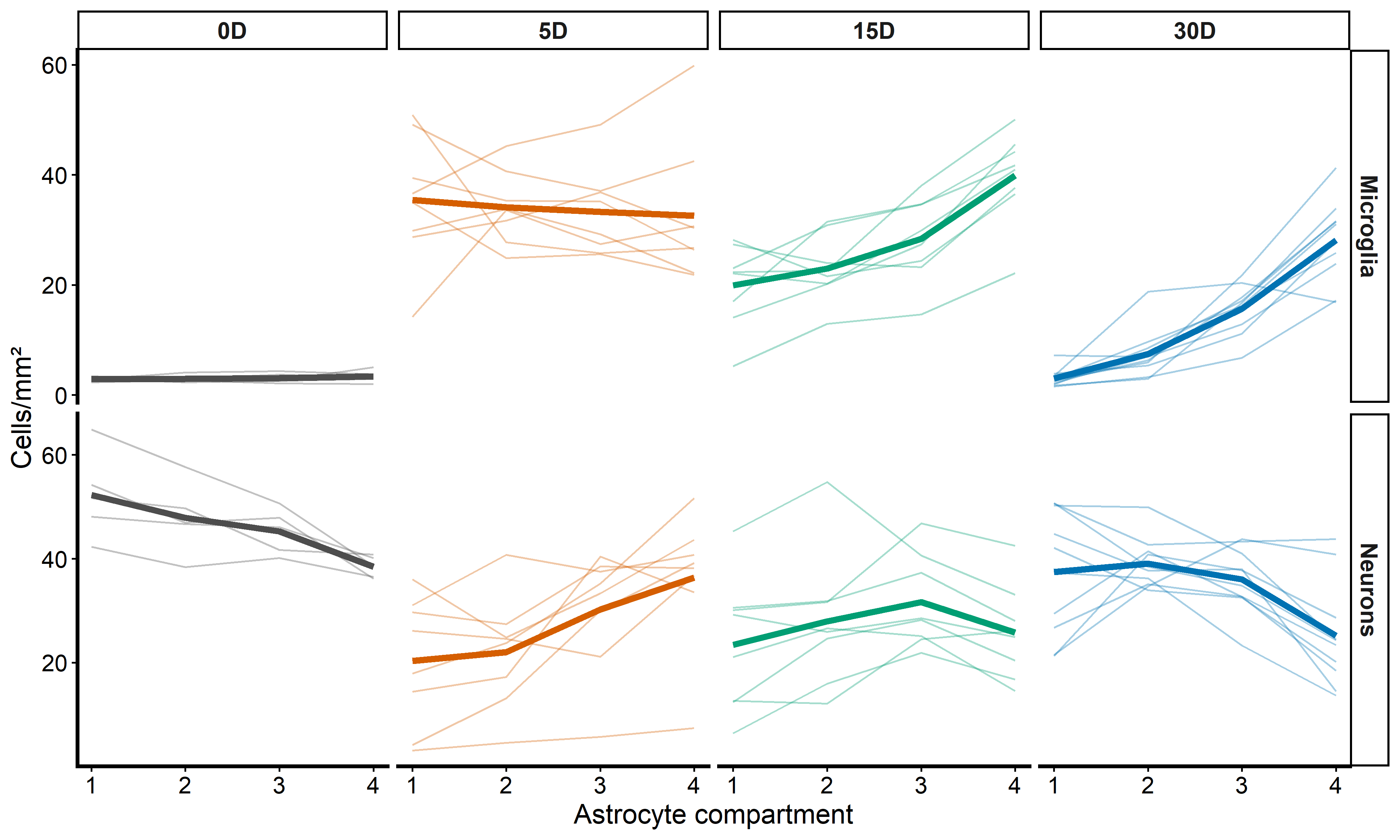
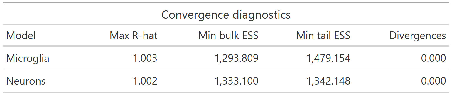
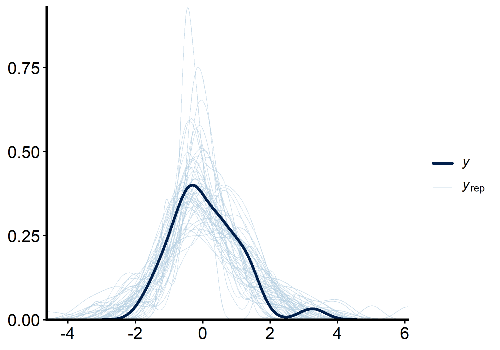
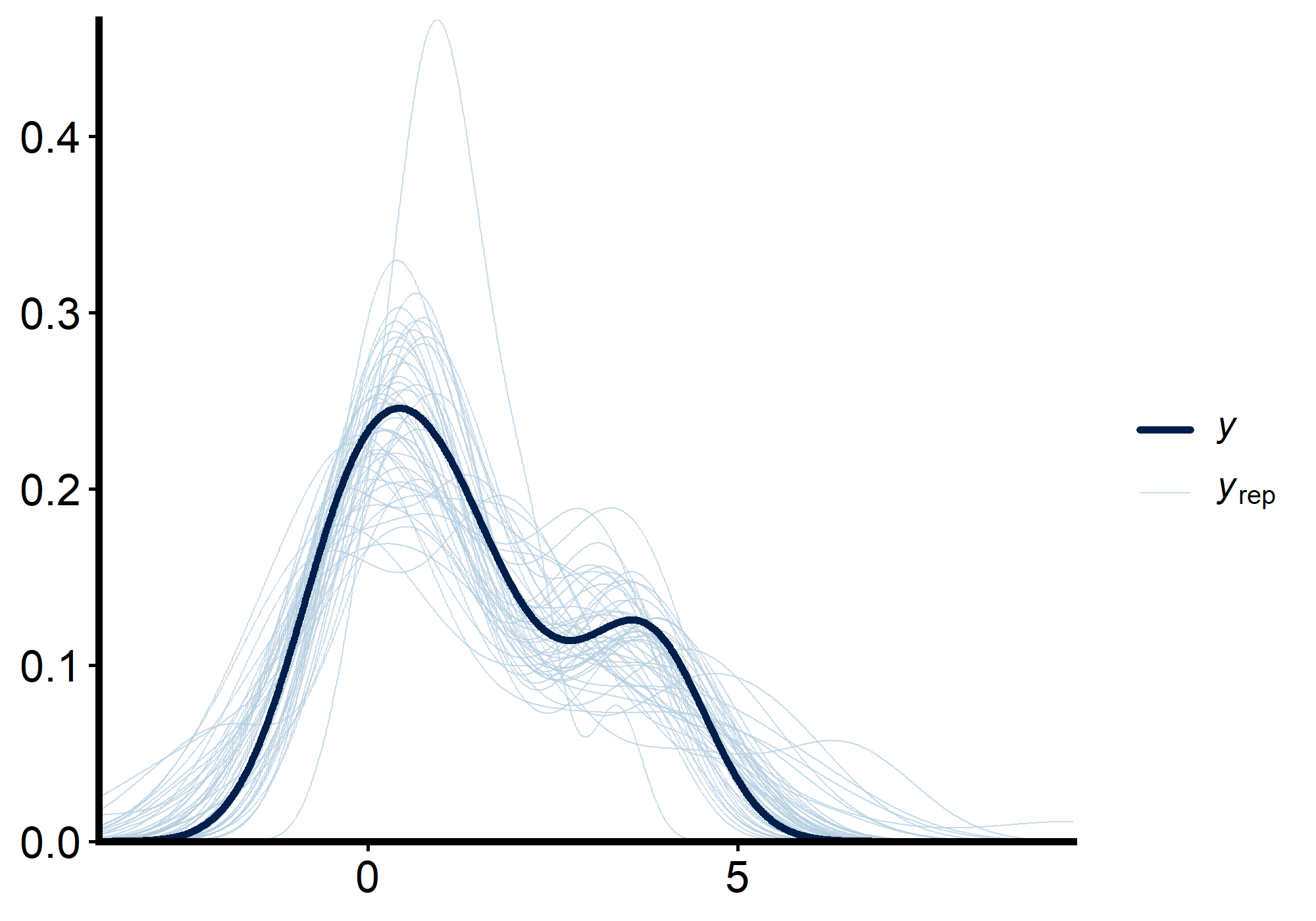
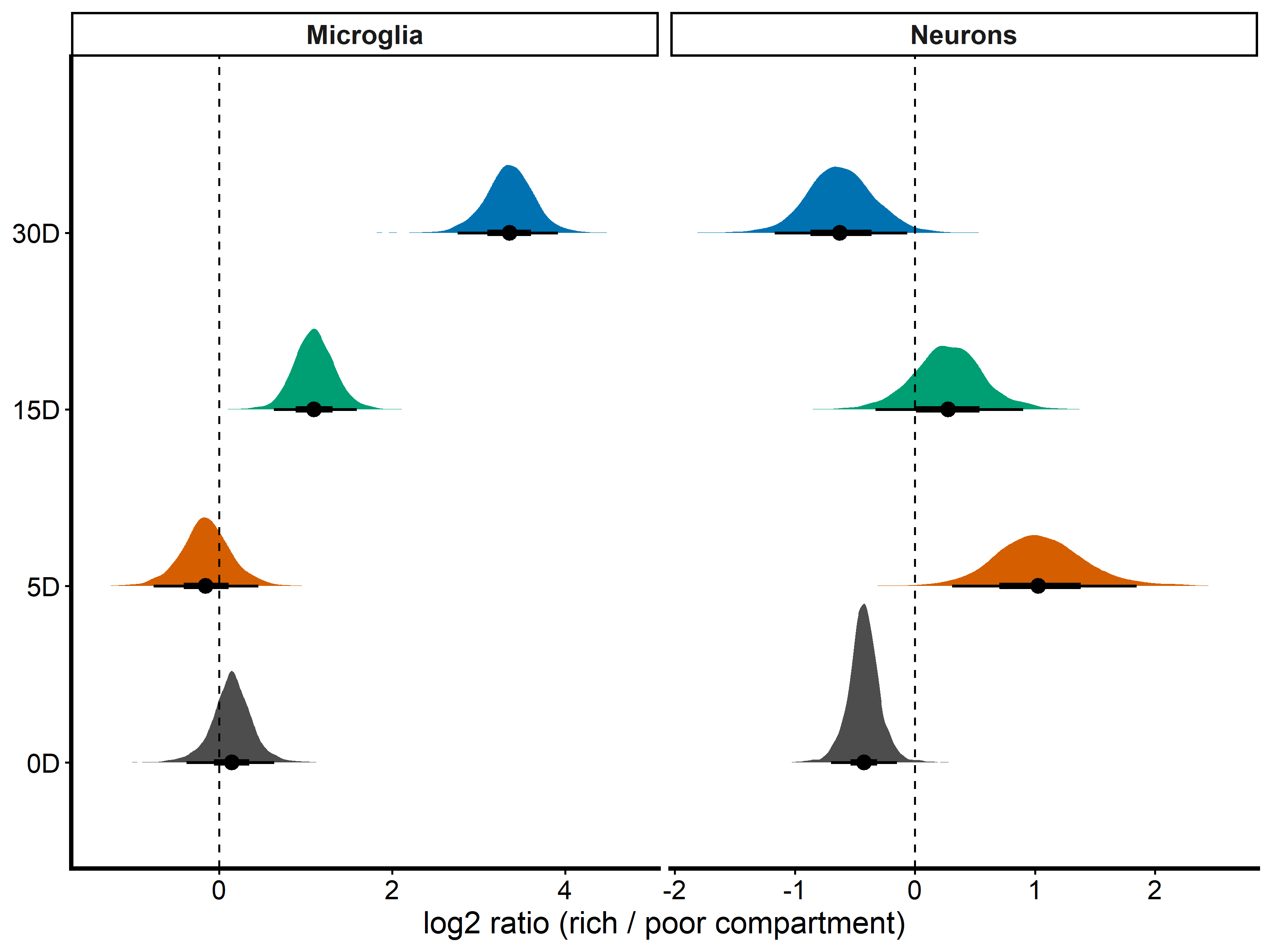
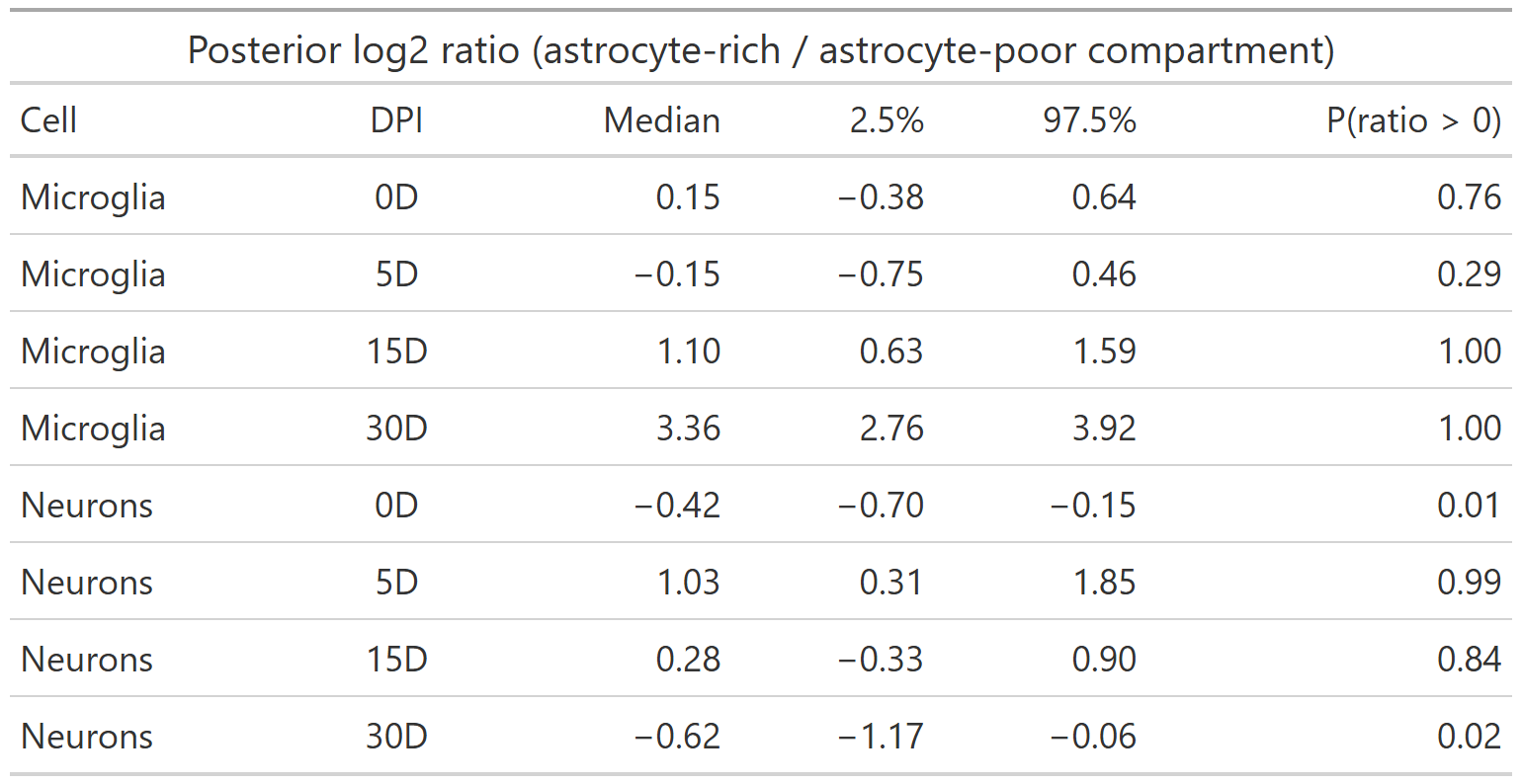
At the end of Part 1 [1] I told you that, in my data, there were fewer neurons where there were more astrocytes. I also left you an exercise: add the days post-ischemia (DPI) to the model and see whether that estimate changes. I had fitted one slope for all the animals, even though a brain at 5 days and a brain at 30 days are not the same tissue.
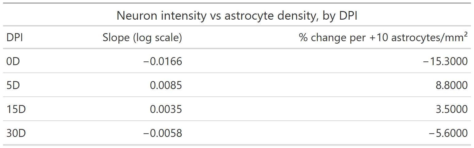
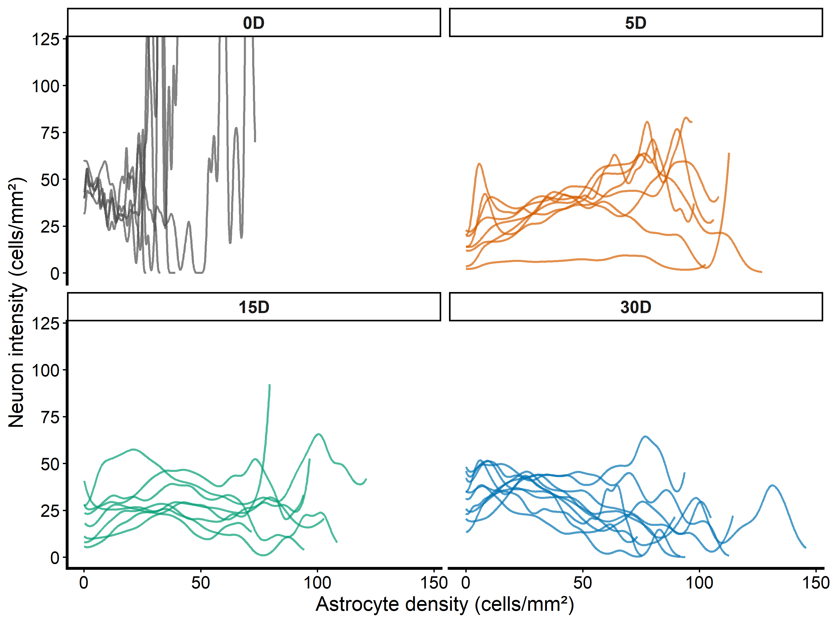
This post is that exercise, done properly. The answer is uncomfortable: the pooled slope I showed you hides a change of sign, and it describes none of the four time points well.
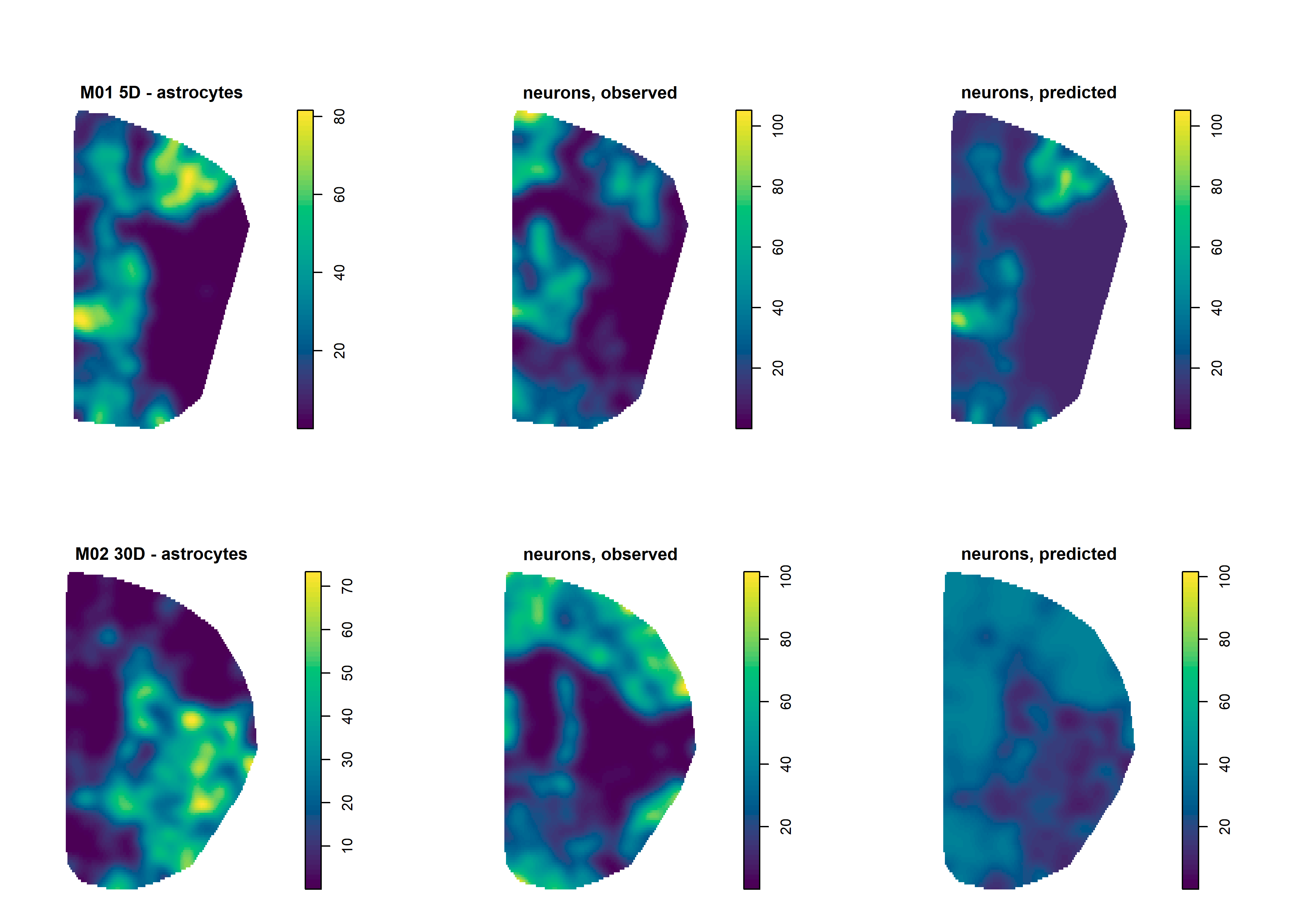
I believe the useful question is not how many cells there are, but how the tissue is compartmentalised, and what the distribution of one cell type tells us about another. For me, the way cells are arranged in the tissue is a key determinant of how they can interact. A cell count cannot see that.…